Atlas of the Neonatal Rat Brain
Book Details
Delivery Location
Delivery fee: Select location
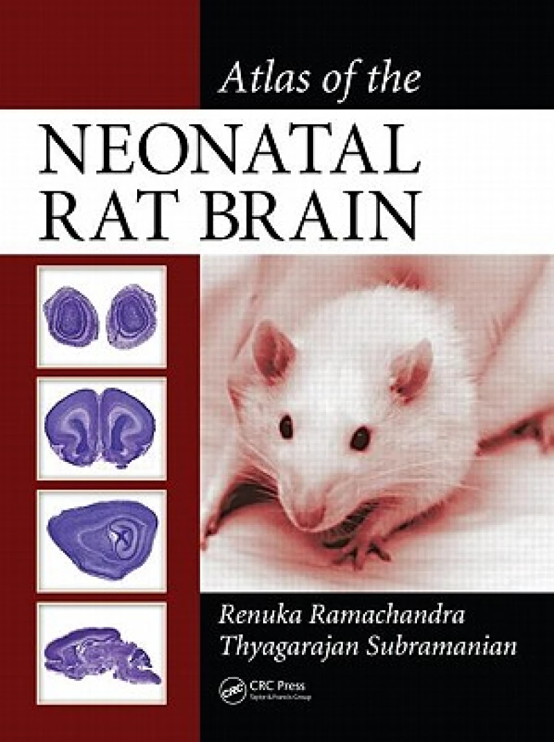
The first histological atlas of the rat neonatal brain, this volume provides researchers in neurology, pharmacology, biology, and veterinary science with detailed color images of the normal developing rat brain. Each plate is a photomicrograph composite that shows the entire brain in sagittal and coronal sections at each stage and stained with cresyl violet. Various brain nuclei and their connections are specifically labeled. Since changes in the rat brain post-birth are so rapid, images are provided for postnatal days one, five, and fifteen.
Atlas of the Neonatal Rat Brain provides photographic, histological illustrations of the anatomical features of the neonatal rat brain at postnatal (P) days P-1, P-7, and P-14.
The sections are Nissl stained with Cresyl violet, creating photomicrographs with high resolution and clarity. The structures are directly labeled on the images, making it easier to correlate data. Additional images are available as electronic resources for individuals who seek images not represented in this volume, and the electronic version allows labels to be removed so the atlas can be used as a teaching tool.
The P-1 section contains 30 coronal plates and 14 sagittal plates and the P-7 section includes 27 coronal plates and 24 sagittal plates. The final P-14 section shows 41 coronal plates and 21 sagittal plates. Each set consists of contiguous sections from individual animals, and selections were based on the structural variability represented.
Get Atlas of the Neonatal Rat Brain by at the best price and quality guaranteed only at Werezi Africa's largest book ecommerce store. The book was published by Taylor & Francis Inc and it has pages.